












14,90 €

Livraison 24/48H en France Métropolitaine - 0,01€ à partir de 35,00€ - 3,95€ jusqu'a 34,99€
Expédition le jour même pour les commandes passées sur les livres "EN STOCK" avant 15h30

Questions, conseils & commandes spécifiques :
La librairie de référence pour toutes les professions médicales et paramédicales
Questions, conseils & commandes spécifiques :
A Paraitre en Imagerie
Dernières parutions Sauramps Médical
Imagerie de la ceinture pelvienne et du membre inférieur
Nouveautés


La Réathlétisation de l'épaule
Dernières parutions Sauramps Médical
Nouveautés Santé Publique
Dernières parutions Sauramps Médical


Le management à la Mayo Clinic
| Auteur(s) | Farhood Saremi |
| Éditeur | THIEME |
| Collection | Imaging anatomy - Thieme |
| Date | 17/06/2026 |
| Pages | 1322 |
| Taille | 21 x 27 |
| Type | Relié |
| Langue | Anglais |
| ISBN | 9781626239869 |
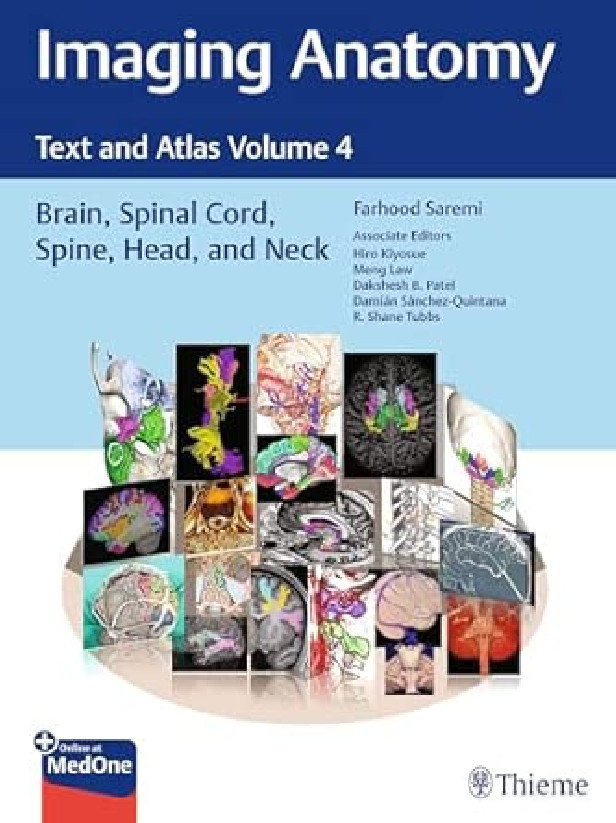
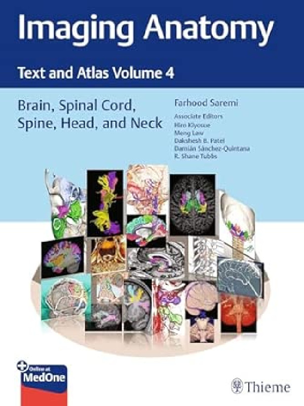
Normal imaging anatomy and variants, including diagnostic and surgical anatomy, are the cornerstones of radiologic knowledge. Imaging Anatomy: Text and Atlas, Volume 4: Brain, Spinal Cord, Spine, Head, and Neck is the fourth in a series of richly illustrated radiologic references edited by distinguished radiologist Farhood Saremi. The atlas is coedited by esteemed colleagues Hiro Kiyosue, Dakshesh Patel, Damian Sanchez-Quintana, and R. Shane Tubbs with contributions from an impressive cadre of international authors. Succinctly written text and superb images provide readers with a virtual, user-friendly dissection experience.
This exquisitely crafted atlas combines fundamental core anatomy principles with modern imaging and post-processing methods to increase understanding of intricate anatomical features. The book features 36 chapters, several of which are available exclusively online.
The chapters cover terminology, classification, and detailed anatomic imaging of the brain, spinal cord, head, and neck structures, along with discussion of relevant associated components for each anatomical area.
This unique resource provides an excellent desk reference for differentiating normal versus pathologic anatomy. It is essential reading for medical students, radiology and neurosurgery residents, neuroradiologists, and neurosurgeons.
DANS LA MÊME COLLECTION :

À PARAÎTRE OU DERNIÈRE PARUTION DANS LA MÊME CATÉGORIE :


Sur commande
Expédition sous 6 à 15 jours
Bénéficiez de la remise de 5% en choisissant le retrait en magasin

d'achats en France Métropolitaine

sous 14 jours ouvrés


Lundi au Vendredi : de 10h à 18h30
au 04 67 63 62 19 ou par email