
















14,90 €

Livraison 24/48H en France Métropolitaine - 0,01€ à partir de 35,00€ - 3,95€ jusqu'a 34,99€
Expédition le jour même pour les commandes passées sur les livres "EN STOCK" avant 15h30

Questions, conseils & commandes spécifiques :
La librairie de référence pour toutes les professions médicales et paramédicales
Questions, conseils & commandes spécifiques :
A Paraitre en Imagerie
Dernières parutions Sauramps Médical
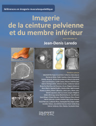
Imagerie de la ceinture pelvienne et du membre inférieur
Nouveautés


La Réathlétisation de l'épaule
Dernières parutions Sauramps Médical
Nouveautés Santé Publique
Dernières parutions Sauramps Médical


Le management à la Mayo Clinic
| Auteur(s) | Surjith Vattoth |
| Éditeur | ELSEVIER HEALTH SCIENCES |
| Date | 26/06/2024 |
| Pages | 664 |
| Taille | 27 X 21 |
| Type | Relié |
| Langue | Anglais |
| ISBN | 9780443249648 |
Written by global leaders in the field, Imaging Anatomy: Head and Neck, second edition, provides a thorough understanding of the detailed normal anatomy that underlies contemporary imaging.
This must-have reference employs a templated, highly formatted design; concise, bulleted text; and state-of- the-art images throughout that identify the clinical entities in each anatomic area, offering a unique opportunity to master the fundamentals of normal anatomy and accurately and efficiently recognize pathologic conditions.
Features hundreds of detailed, full-color illustrations and more than 900 high-resolution, cross-sectional radiologic images that together illustrate the fine points of imaging anatomy for new and experienced head and neck imaging specialists
Contains new chapters on external nose anatomy, the facial nerve in temporal bone, minor fissures and sutures around the temporal bone, and temporal bone anatomy on photon-counting detector (PCD) CT
Provides updated, enlarged images and captions in areas such as facial muscles and the superficial musculoaponeurotic system, and frontal recess and related air cells
Includes extensive new content on PCD CT; new details on relatively unknown anatomical foramina, such as the vomerovaginal canal and canaliculus innominatus; new content based on the International Frontal Sinus Anatomy Classification; and minute details on the course of nerves in the head and neck
Includes a series of successive imaging slices in each standard plane of imaging (coronal, sagittal, and axial) to provide multiple views that further support learning
Depicts common anatomic variants and covers the common pathological processes that manifest with alterations of normal anatomic landmarks
Reflects new understandings of anatomy due to ongoing anatomic research as well as new, advanced imaging techniques
Presents essential text in an easy-to-digest, bulleted format, enabling imaging specialists to find quick answers to anatomy questions encountered in daily practice
Includes an eBook version that enables you to access all text, figures, and references with the ability to search, customize your content, make notes and highlights, and have content read aloud. Any additional digital ancillary content may publish up to 6 weeks following the publication date
Surjith Vattoth : Professor of Diagnostic Radiology & Nuclear Medicine, Division of Neuroradiology, Rush University Medical Center, Chicago, Illinois, USA
À PARAÎTRE OU DERNIÈRE PARUTION DANS LA MÊME CATÉGORIE :

.
Du même auteur :
Sur commande
Expédition sous 20 à 30 jours - Les éditions Elsevier Health Sciences sont en reorganisation logistique du 25/09 au 15/10. Cela explique l'allongement du délai d'expédition pour cet éditeur sur notre site. Merci de votre compréhension.
Bénéficiez de la remise de 5% en choisissant le retrait en magasin

d'achats en France Métropolitaine

sous 14 jours ouvrés


Lundi au Vendredi : de 10h à 18h30
au 04 67 63 62 19 ou par email


